FIG 3.
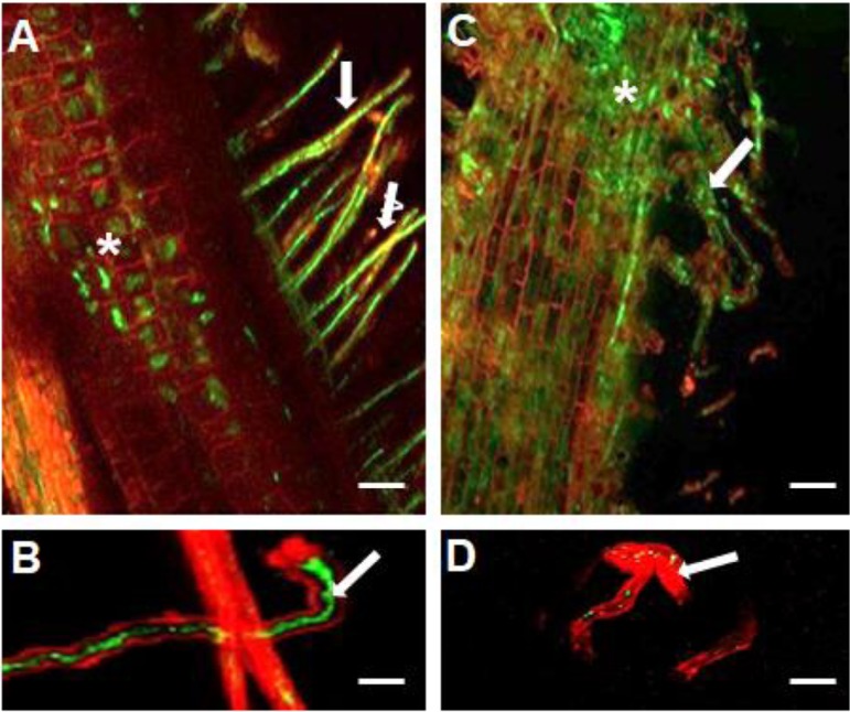
Confocal laser scanning microscopy of colonization and root hair infection of soybean by GFP-marked USDA110 WT and ΔssuD mutant strains. (A) The WT strain colonizing the root hairs (arrows) and central root tissues (*). (B) Root hair infection thread formed by the WT strain. (C) The ΔssuD mutant showed diffuse colonization of root hairs (arrows) and the main root surface (*). (D) A few GFP-expressing ΔssuD mutant bacteria can be seen within the infected root hair (arrow). Bars, 100 μm for A and C, 30 μm for B and D.

