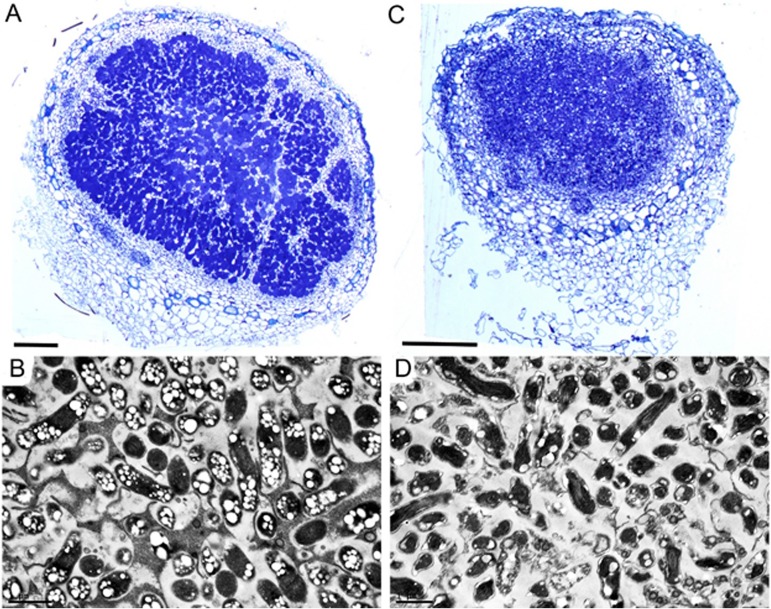
FIG 4.
Microscopic analysis of soybean nodules formed by USDA110 and the ΔssuD mutant strains. (A) Light micrograph of a transverse section of a nodule formed by the WT strain showing intense toluidine blue staining of bacteria. (B) Transmission electron micrograph (TEM) of WT nodule showing elongated bacteroids containing polyhydroxybutyrate granules in symbiosomes. (C) Light micrograph of nodule formed by the ΔssuD mutant showing diffuse toluidine blue staining. (D) TEM of a nodule formed by the ΔssuD mutant showing degraded bacteroids. Bars, 200 μm.

