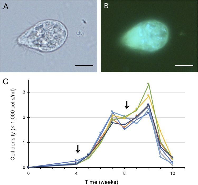
FIG 1.
An anaerobic scuticociliate strain, GW7. (A and B) Phase-contrast micrograph (A) and fluorescence micrograph (B) of a cell of strain GW7. Autofluorescence of coenzyme F420 (light blue) was detected. (C) Growth curve of strain GW7. The timing of supplementation of the food bacterium (0.05 g) is indicated by an arrow. Different-colored lines show each replicate (n = 7). Bars, 10 μm (A and B).

