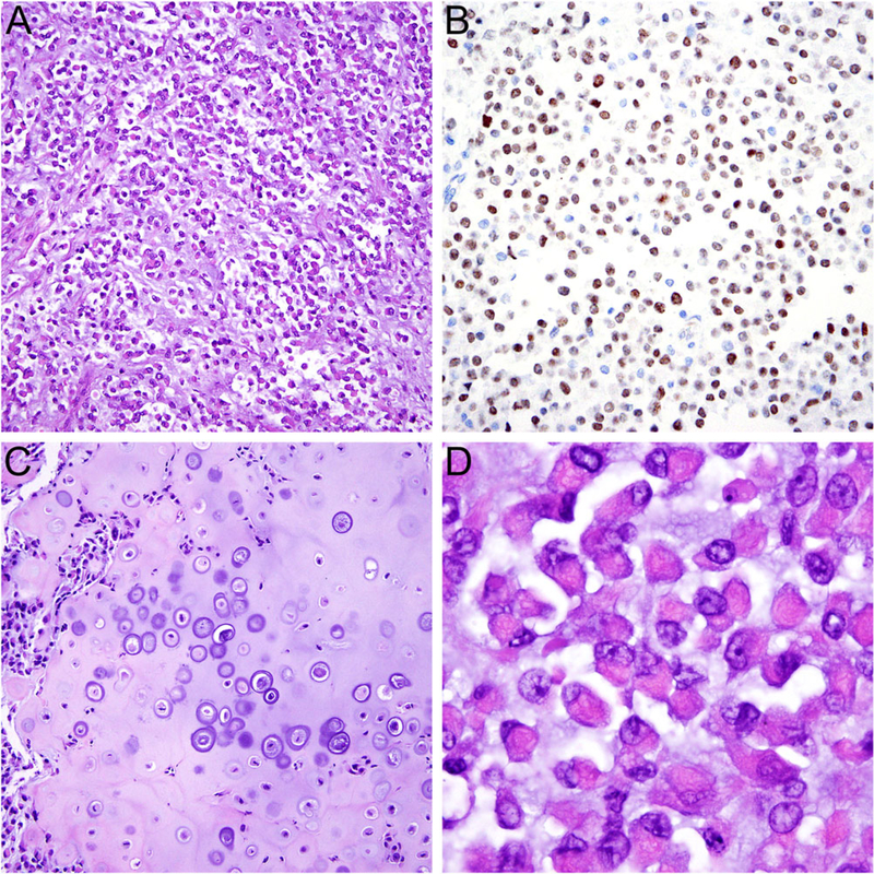
FIGURE 4.
Histologic features of NUT variant with NSD3-NUTM1 fusion. A, The tumor consisted of round to ovoid cells mostly arranged in variably cohesive sheets embedded in a myxoid stroma. B, The tumor cells showed diffuse nuclear expression of NUT. C, Focal heterologous cartilage formation and D, areas with prominent rhabdoid cytomorphology were present, resembling features of myoepithelial carcinoma

