Figure 1.

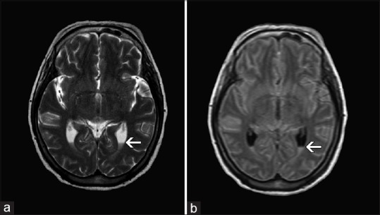
Magnetic resonance imaging of brain T2W image (a), FLAIR image (b) showing intra ventricular hyperintensity and fluid level in the dependent portion of occipital horn of bilateral lateral ventricle

Magnetic resonance imaging of brain T2W image (a), FLAIR image (b) showing intra ventricular hyperintensity and fluid level in the dependent portion of occipital horn of bilateral lateral ventricle