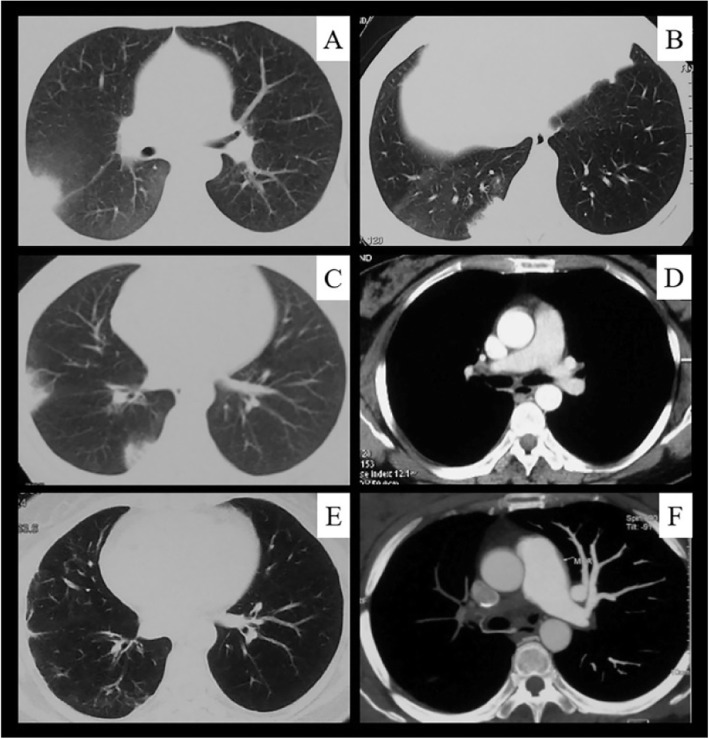
Fig. 4.
Serial computed tomography (CT) images in a 40-year-old woman with Takayasu’s arteritis. The patient was admitted to our hospital because of shortness of breath. She had developed recurrent chest and back pain and hemoptysis 4 years previously. a–c, CT images show recurrent subpleural wedge-shaped opacities during the initial 6 months after disease onset. d CT pulmonary angiography (CTPA) image obtained at the same time as in (c) showed right pulmonary artery stenosis. e, four years later, a CT image shows peripheral scarring from previous infarcts. f, CTPA image obtained at the same time showed right pulmonary artery occlusion

