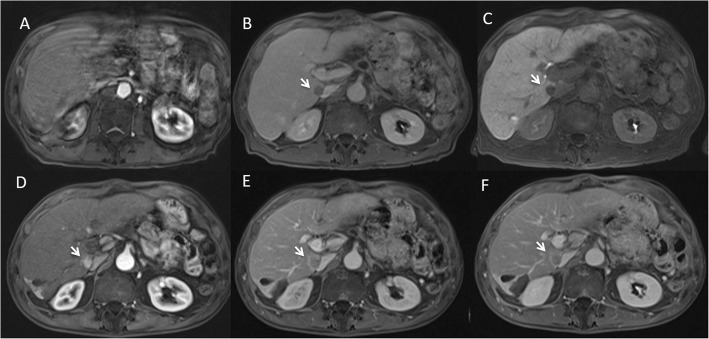
Fig. 1.
Man 73 years with HCC nodule. In a, b and c GD-EOB-DTPA M0 study. In a arterial phase (VIBE T1-W FS), the HCC located to VII segment is not detect and it is due to lower quality of this phase. In b (late phase of contrast study) and c (HPB phase of contrast study) the nodule is detected (arrows). In D, E and F Gd-BT-DO3A M3 study. In d (arterial phase) the HCC nodule (arrow) shows wash-in with wash-out and capsule appearance during portal (e) and equilibrium (f) phase of contrast study

