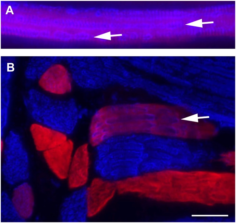
Figure 6.
Longitudinal sections of 6-week-old rat labeled for type I (blue) or type IIA (red) fibers. Hybrid fibers are seen as purple. Closer examination of staining patterns within the hybrids shows that individual myofibrils comprised of type I MHC are distinct within single hybrid fibers (arrows). Sarcomeric banding is clearly seen within myofibrils in A. Figs A and B are different regions of the same muscle. Scale bar = 25 µm.

