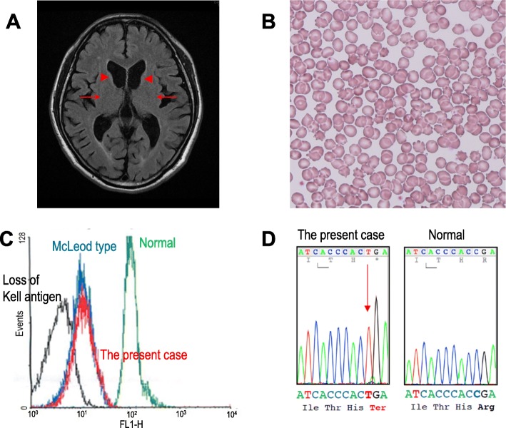
Fig. 1.
a MRI fluid-attenuated inversion recovery axial image shows atrophy of the caudate nuclei (arrowheads) and putamen (arrows) bilaterally. b Erythrocyte acanthocytosis present in a peripheral blood smear. c Flow cytometry revealed reduced Kell red blood cell antigens in the peripheral blood (red peak indicates the present case). d Sequencing of the XK gene disclosed mutation c.397C > T (p.Arg133Ter) in exon 2

