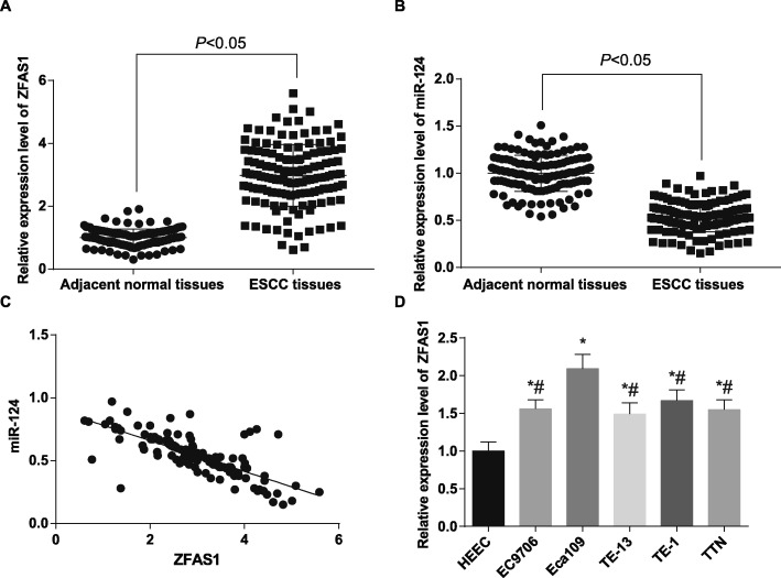
Fig. 1.
ZFAS1 is highly expressed and miR-124 is lowly expressed in ESCC. a: Detection of ZFAS1 expression in ESCC tissues and their adjacent normal tissues by RT-qPCR (n = 136). b: Detection of miR-124 expression in ESCC tissues and their adjacent normal tissues by RT-qPCR (n = 136). c: Correlation between ZFAS1 and miR-124 expression in ESCC tissues analyzed by Pearson correlation analysis. d: RT-qPCR detected ZFAS1 expression in human normal esophageal epithelial cells HEEC and five ESCC cell lines. * p < 0.05 vs. HEEC cells. # p < 0.05 vs. Eca109 cells. Measurement data were depicted as mean ± standard deviation, comparisons between two groups were conducted by independent sample t-test, and comparisons among multiple groups were assessed by one-way analysis of variance followed by Tukey’s post hoc test. Repetitions = 3 in cellular experiment

