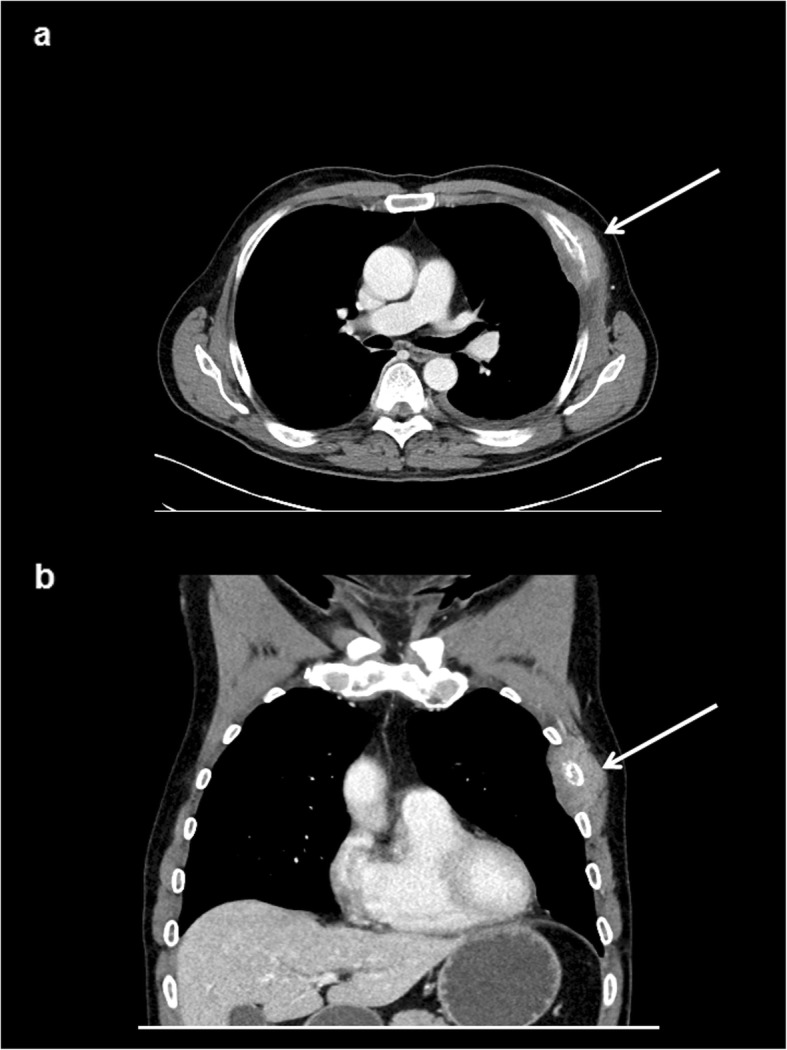
Fig. 1.

Myeloid sarcoma of the chest wall in a male patient with known acute myeloid leukemia in a relapse setting. Contrast enhanced computed tomography in axial (a) and coronal plane (b) showing a relatively homogenous of the third left rip with an infiltration of the adjacent intercostal muscles (arrows)
