
Figure 3.
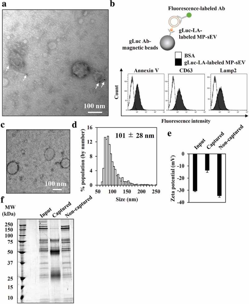
Characterization of gLuc-LA-labelled MP-sEVs (gLuc-LAMP-sEVs). (a) Transmission electron microscopy (TEM) observation of gLuc-LAMP-sEVs stained with protein A-gold nanoparticles (indicated by arrows) after reacting with an anti-gLuc antibody. (b–f) gLuc-LAMP-sEVs in the SEC eluate sample was immunocaptured by gLuc antibody-coated magnetic beads. (b) To confirm the sEV capturing by the beads, the sEVs-beads complexes were subsequently stained with the indicated FITC-annexin V (high affinity to PS), PE-anti-CD63 antibody, or alexa fluor 488-anti-Lamp2 antibody and analysed by flow cytometry. BSA was set as a control sample against the sEV. Then, the sEV was eluted from the beads and psychochemical properties as well as protein composition was identified as follow: (c) sEV morphology by TEM analysis, (d) Size histogram measured by qNano instrument, (e) Zeta potential of sEV, and (f) SDS-PAGE analysis (0.7 µg/lane). The input and non-captured fraction of the immunocapturing was simultaneously analysed for zeta potential and SDS-PAGE.