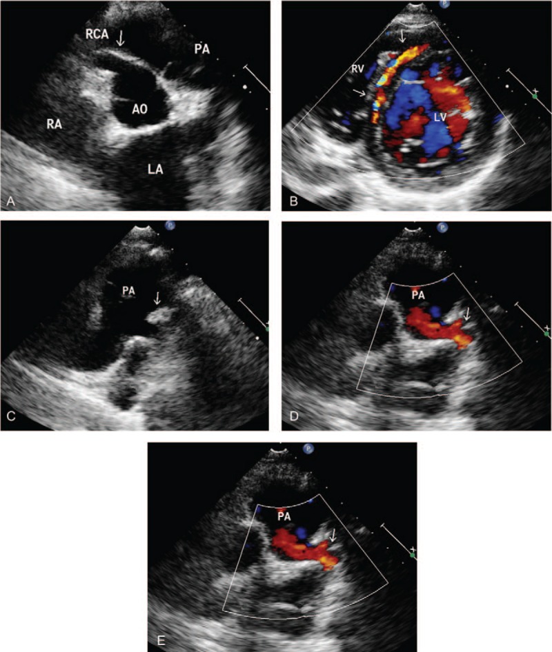
Figure 1.

ALCAPA in a 8-year-old male. (A) Transthoracic echocardiography shows a dilated right coronary artery (arrows). (B) Color Doppler flow image reveals the collateral vessel in the interventricular septum (arrow). (C) Parasternal short-axis view shows the left coronary artery originating from pulmonary artery (arrow). (D) Color Doppler reveals a retrograde flow from the ostium of the LCA to the PA (arrow). (E) Pulse Doppler showed the retrograde red shunt a lowvelocity, prominently diastolic flow spectrum. LA = left atrium, LV = left ventricle, RA = right atrium, AO = aortic, PA = pulmonary artery, RCA = right coronary artery.
