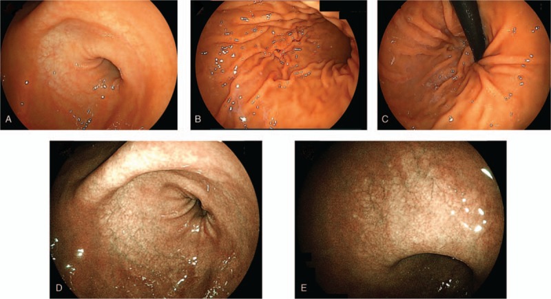
Figure 3.

Esophagogastroduodenoscopy revealed gastric mucosal atrophy localized from the antrum to the upper corner. The atrophic grade was C-2 according to the Kimura–Takemoto classification system. The esophagus and duodenum did not present any abnormality. Linked color imaging findings for (A) antral zone, (B) gastric body, (C) cardia and fornix, (D) antral zone, (E) gastric angle.
