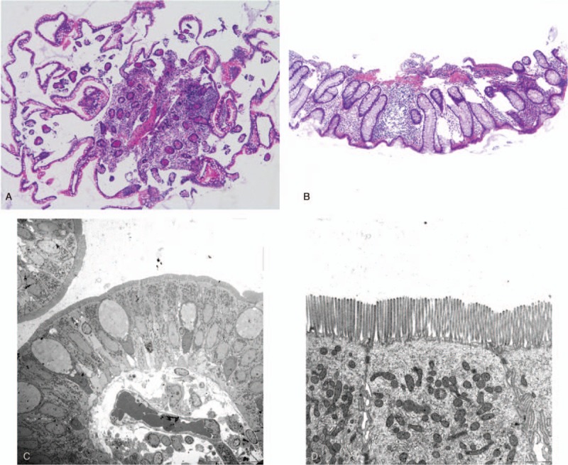
Figure 6.

(A, B) The only pathological findings of the terminal ileum and rectum mucosa were mild edema and infiltration of inflammatory cells in the lamina propria (HE stain). (C, D) The electronic microscopic images showed no abnormal villi, similar to that for the duodenum. HE = hematoxylin and eosin.
