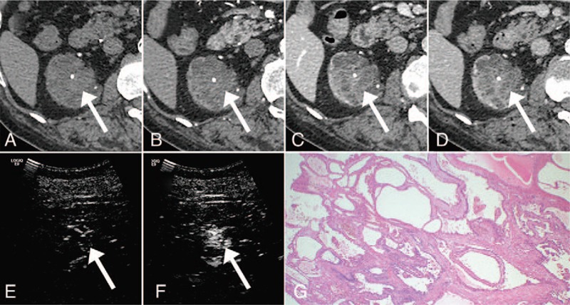
Figure 3.

A 65-year-old male with multilocular cystic renal cell carcinoma (RCC). A, Unenhanced computed tomography (CT). B, Dynamic contrast-enhanced CT, early phase. C, Dynamic contrast-enhanced CT, parenchymal phase. D, Dynamic contrast-enhanced CT, delayed phase. E, Precontrast ultrasonography. F, Ultrasonography 30 seconds after the administration of contrast medium. The enhancement was ambiguous on the dynamic contrast-enhanced CT, while it was obvious on the contrast-enhanced US. G, Hematoxylin and eosin-stained slide. Multiple fibrous septa are lined by a single layer of low-grade tumor cells.
