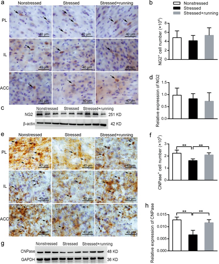
Fig. 4. The effects of running exercise on the total NG2+ cell numbers and the total CNPase+ cell number in the mPFC of CUS rats.
a Representative pictures of immunohistochemical staining with anti-NG2 antibody in each subregion of the mPFC of the nonstressed rats, stressed rats and stressed + running rats. Scale bar = 40 μm. b Total NG2+ cell number in the mPFC in the nonstressed group, stressed group and stressed + running group (mean ± SD, n = 5 per group). c The protein expression of NG2 in the mPFC of each group rats was detected using western blot. d Semiquantitative analyses of the protein level of NG2 in the mPFC from each group (mean ± SD, n = 8 per group). e Representative pictures of immunohistochemical staining with anti-CNPase antibody in each subregion of the mPFC from the nonstressed rats, stressed rats and stressed + running rats. Scale bar = 40 μm. f Total CNPase+ cell number of mPFC in the nonstressed rats, stressed rats and stressed + running rats (mean ± SD, n = 5 per group). g The protein expression of CNPase in the mPFC of each group of rats was detected using western blot. h Semiquantitative analyses of the protein level of CNPase in the mPFC from each group (mean ± SD, n = 8 per group). **p < 0.01. PL prelimbic, IL infralimbic, ACC anterior cingulate cortices.

