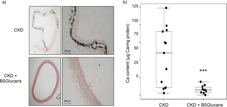
Figure 4.
Anti-calcifying protection by dietary BßGlucans. (a) Representative calcium deposition measured by Von Kossa (black) staining (Inset bars indicate relative scale) in thoracic aortas from 5/6NX rats fed a high P diet with none (CKD; n = 13) or 2 mg of Bßglucans/g diet (CKD + Bßglucans; n = 13) during 4 weeks. (b) Quantification of calcium deposition in thoracic aortas described in (a); boxplot analysis of changes in calcium content, *p < 0.05 vs. CKD.

