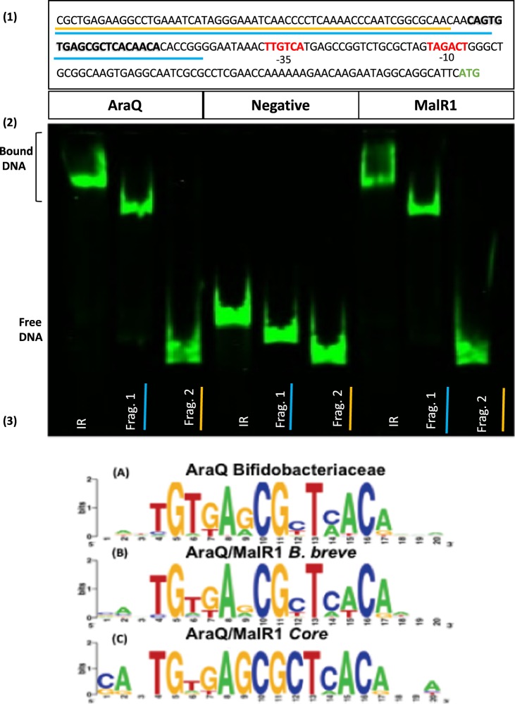
Figure 1.
Fragmentation analysis and AraQ/MalR1 binding. Panel (1) depicts the promoter region of pyk (Bbr_0757) used for fragmentation analysis. −35 and −10 sites are indicated in red and the ATG start codon is indicated in green. The TFBS is indicated in bold. The fragment including the TFBS (Frag. 1) is underlined in blue, the fragment excluding the TFBS (Frag. 2) is underlined in yellow, while the full fragment is referred to as IR. Panel (2) EMSA to investigate AraQ and MalR1 abilities to bind to Bbr_0757 promoter region fragments (IR, Frag 1 and Frag 2). All reactions contain 0.5 nM Ird labelled DNA and 150 nM of either AraQ or MalR1 protein, while negative reactions contain 0 nM protein. Uncropped gel image available in Supplemental Fig. S5. Panel (3), (A) Illustrates consensus AraQ/MalR1 binding motif across the Bifidobacteriaceae family (B) Depicts the binding motif of AraQ/MalR1 in B. breve. (C) Depicts the AraQ/MalR1 binding motif for core members of the regulon.

