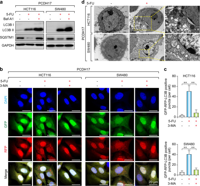
Fig. 3.
PCDH17 increases autophagosome formation and autophagic flux in CRC cells. a PCDH17-transfected HCT116 and SW480 cells were treated with 20 μM 5-FU with or without 10 μM BafA1 for 24 h, and the protein levels of LC3B and SQSTM1 were assessed by western blotting. b HCT116/PCDH17 and SW480/PCDH17 cells were transfected with the GFP-RFP-LC3 plasmid overnight and transferred onto coverslips. After exposure to 20 μM 5-FU with or without 3-MA for 24 h, representative images of LC3-II-positive puncta were obtained with a confocal fluorescence microscope. c The quantitative analyses of the number of fluorescent puncta are shown. The experiments were performed in triplicate. **p < 0.01. d Electron microscopy shows the ultrastructures of autophagosome and autolysosome vesicles in these cells. The experiments were performed in triplicate. Bar = 2 μm.

