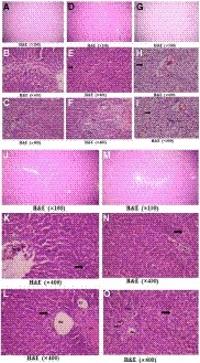
Figure 5.

Photomicrographs of H&E‐stained sections of rat livers, (a–c) the control group showing the normal hexagonal or pentagonal liver lobules (a). Hepatocytes were arranged in plates one cell thick, having acidophilic cytoplasm with one or occasionally two spherical nuclei with prominent nucleoli (b). (c) Portal triad containing a branch of hepatic artery (HA), portal vein (PV), and a small bile duct (BD). The phagocytic Kupffer's cells adhered to the endothelial lining of hepatic blood sinusoids (*). (d–f) the hyperuricemic group had slightly disturbed hepatic architecture (arrow in e). (f) Showed features of portal inflammation in the form of dilated congested blood vessels (HA and PV) and mononuclear cellular infiltration (* in f). (g–h) Fructose‐supplemented hyperuricemic group showing extensive hepatic damage, congested central, and portal veins (g). Many necrotic areas with complete destruction of hepatocytes were present as more vacuolated foamy cytoplasm with darkly stained pyknotic nuclei (arrow in h and i), being more at the periphery of the hepatic lobules. (j–k) l‐Carnitine‐treated hyperuricemic group showing nearly normal hepatic architecture, but some hepatocytes were still having vacuolated cytoplasm (arrow) and normal vesicular nuclei (k, l) with little leukocytic infiltration at the portal triad (l). (m‐o) l‐Carnitine‐treated fructose‐supplemented hyperuricemic group showing apparently normal hepatic architecture, despite the presence of more hepatocytes with vacuolated cytoplasm (arrow in n and o) but with normal vesicular nuclei (n) and with little leukocytic infiltration at the portal triad (o)
