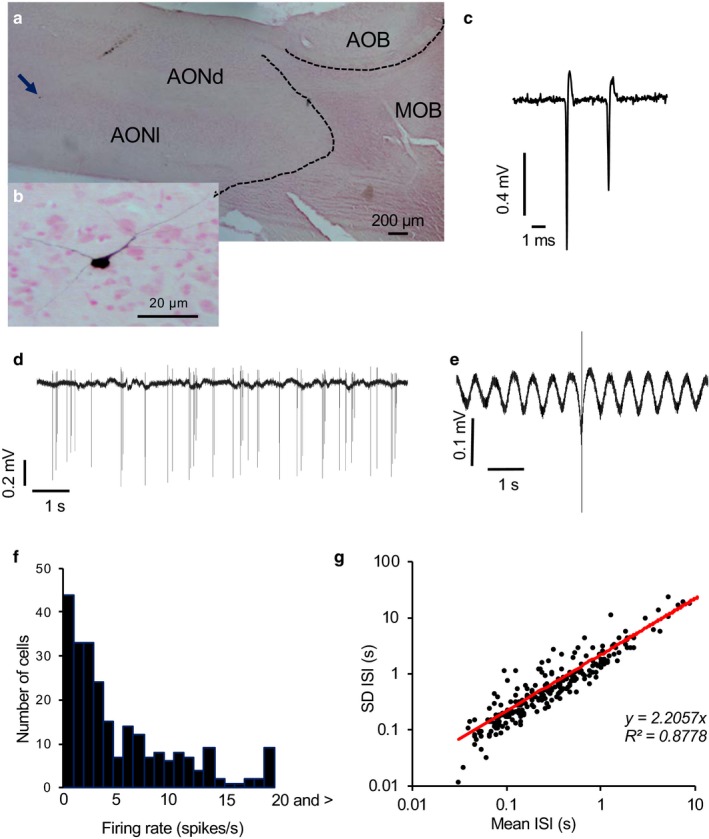
Figure 1.

Juxtacellularly labeled neuron in the AON. (a) Juxtacellularly labeled neuron (arrowhead) in the rat AON shown at higher magnification on the lower. AOB; accessory olfactory bulb, MOB; main olfactory bulb, AONd; anterior olfactory nucleus dorsal, AONl; anterior olfactory nucleus lateral. (b) Higher magnification of the neuron arrowed in A showing multipolar neuron with oval cell body. (c) Extract of voltage trace showing individual spikes. This neuron showed decreasing spike height in spikes clustered at high frequency. (d) Voltage record spontaneous activity pattern showing rhythmic discharge of spikes, including short bursts. (e) Average action potential from this neuron (average of all spikes fired during 300 s) expanded to show rhythmic oscillations of voltage. (f) Distribution of spontaneous firing rates of a sample of 240 AON neurons. (g) The SD of ISIs is closely correlated with the mean ISI (R 2 = .88)
