
Fig. 1.
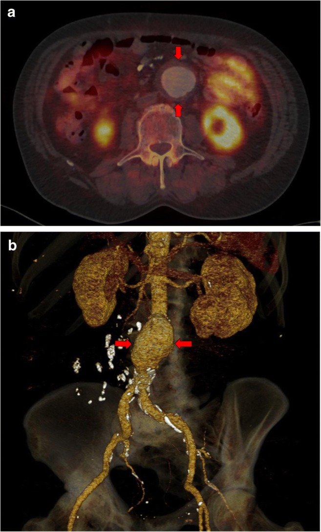
Staging PET/CT examination of patient with prostate cancer. a The subrenal AAA with a diameter of 55 mm (red arrows) visualized by the fusion PET/CT image. b The same patients with the subrenal AAA with a diameter of 55 mm (red arrows) visualized by three-dimensional CT angiography used in stent graft planning