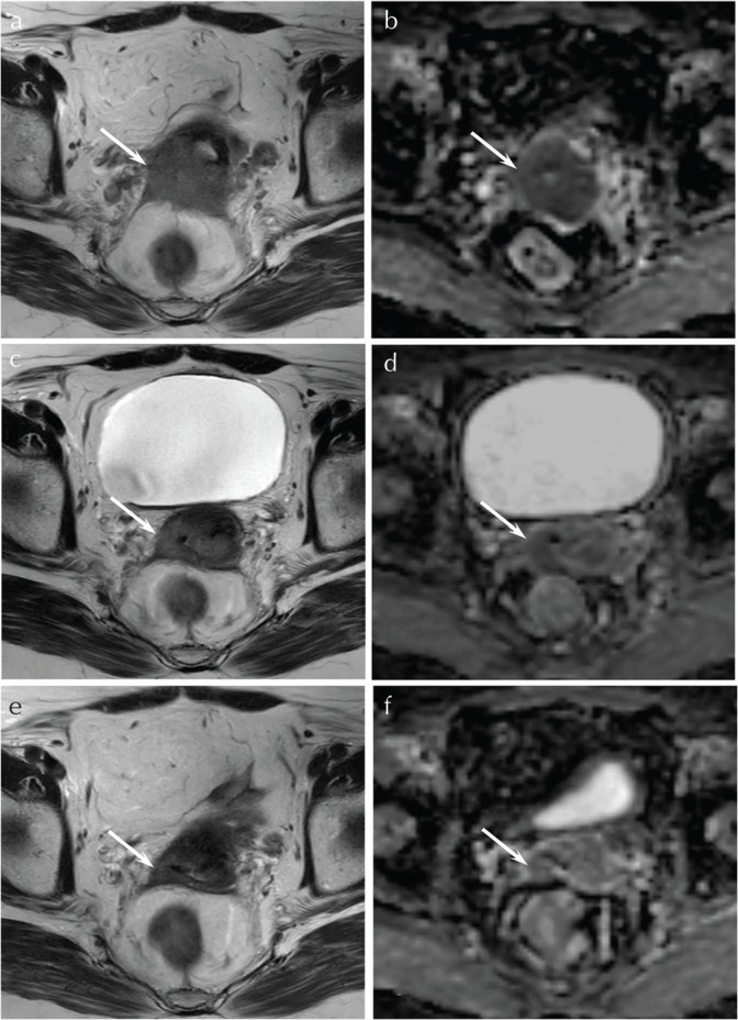
Fig. 1.

A 62-year-old woman with squamous cell carcinoma of the uterine cervix (CR group). (a) T2-weighted fast spin-echo image (TR/TE, 5216/100 ms) before CRT shows an ill-demarcated lesion with parametrial invasion (arrow). (b) ADC map before CRT shows low ADC value (0.85 × 10⁻³ mm2/s) (arrow). (c) T2-weighted fast spin-echo image (TR/TE, 5216/100 ms) during CRT at the dose of 20 Gy shows a decrease in size of the tumor (arrow). (d) ADC map during CRT at the dose of 20 Gy shows an increase in ADC value (1.31 × 10⁻³ mm2/s) with high Δ%ADC20 Gy (55.1%) (arrow). (e) T2-weighted fast spin-echo image (TR/TE, 5216/100 ms) during CRT at the dose of 40 Gy shows a further decrease in size of the tumor (arrow). (f) ADC map during CRT at the dose of 40 Gy shows a further increase in ADC value (1.53 × 10⁻³ mm2/s) with high Δ%ADC40 Gy (80.6%) (arrow). ADC, apparent diffusion coefficient; CR, complete remission; CRT, chemoradiotherapy
