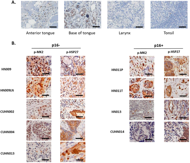
Figure 1.
Phosphorylated MK2 (p-MK2) is overexpressed in head and neck tumor tissue compared to non-cancerous human tissue. A, Normal human head and neck tissue was obtained from the UNM Human Tissue Repository (HTR) via the IRB-approved study, INST 1310 (SRC 008-17). Standard immunohistochemistry was performed on 4-micron cut tissue sections staining for p-MK2. B, Primary head and neck cancer tissue obtained through institutional HTR using two different IRB approved protocols (University of New Mexico: HN009, HN009LN, HN011P, HN011T, HN013; University of Colorado Denver: CUHN002, CUHN004, CUHN0013, CUHN014) were cut and stained for p-MK2. Both p16 negative (p16−) and p16 positive (p16+) primary human HNSCC tissues were examined. Black bar denotes 50 μm size.

