Figure 2.
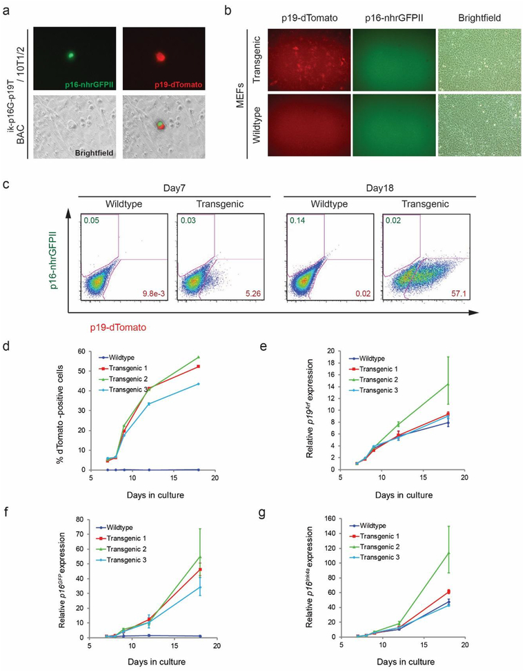
Induction of the Cdkn2a reporter with serial passage of transgenic mouse embryonic fibroblasts. a Fluorescence photomicrograph of 10T1/2 fibroblasts transfected with of BACik-p16G-p19T reporter shows isolated cell expressing both green (p16-nhrGFPII) and red (p19-dTomato) reporters. b, c Fluorescence photomicrograph (b) and flow cytometry plots (c) of MEFs derived from wildtype and BACik-p16G-p19T transgenic mice at passage number 5 (b) or following 7 or 18 days in culture (c). Numerous dTomato positive cells were detected in transgenic cells, but nhrGFPII protein was not visible, even though mRNA was induced (f, g). d Quantitative analysis of dTomato-positive MEFs from three different transgenic lines or a single wildtype line. Cells were quantified by flow cytometry with serial passage for the indicated number of days. e-g Quantitative analysis of relative mRNA expression of endogenous mRNA for Arf (e), p16-nhrGFPII (f), and Ink4a mRNA (g) in wildtype or transgenic MEFs upon serial passage.

