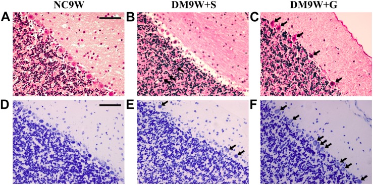
FIGURE 2.
Effects of gastrodin on DM-induced pathological changes in the cerebellar cortex. H&E staining (A–C) and Nissl staining (D–F) of the cerebellar cortex in NC9W, DM9W + S, and DM9W + G groups. Black arrow indicates Purkinje cells. Note the drastic reduction of Purkinje neurons in the DM9W + S group. Note also the wide interstitial spaces around the Purkinje cells in the same group. In DM9W + G, the incidence of Purkinje cells is increased; moreover, the neuropil is now more compact. Magnification: ×400. Bar = 50 μm.

