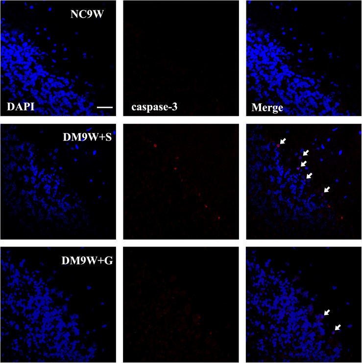
FIGURE 3.
Representative photomicrographs of DAPI (blue) and caspase-3 (red) double staining of the cerebellum in the NC9W, DM9W + S, and DM9W + G groups. Note the increase in incidence of caspase-3 positive cells in DM9W + S (single arrow) compared with the NC9W group. However, in the DM9W + G group, caspase 3 + cells are hardly encountered. Magnification: ×600. Bar = 20 μm.

