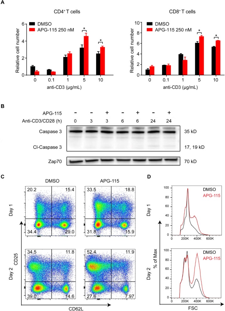
Fig. 2.
APG-115 increase mouse T cell proliferation and enhances mouse CD4+ T cell activation. a CD4+ T and CD8+ T cells were positively selected from mouse spleens using magnetic beads and then stimulated with indicated concentrations of plate-bound anti-CD3 and 2 μg/mL anti-CD28 in the presence of 250 nM APG-115 or DMSO. After 72 h, relative cell numbers were determined using CellTiter-Glo luminescent cell viability assay (Promega) and normalized to unstimulated cultures treated with DMSO control. * P < 0.05. b immunoblots for the expression of caspase 3, cleaved caspase 3, and Zap-70 (loading control) in total cell lysates of anti-CD3/CD28-stimulated CD4+ T cells exposed to APG-115 or solvent control DMSO for 3, 6, or 24 h (h). c CD4+ T cells were positively selected from mouse spleens using magnetic beads and then stimulated with 10 μg/mL plate-bound anti-CD3 and 2 μg/mL anti-CD28 in the presence of 250 nM APG-115 or DMSO for the indicated periods of time. T cell activation markers (CD25 and CD62L) were determined by flow cytometry. CD25high CD62Llow T cells represented an activated population. d an increase in cell size was shown after APG-115 treatment

