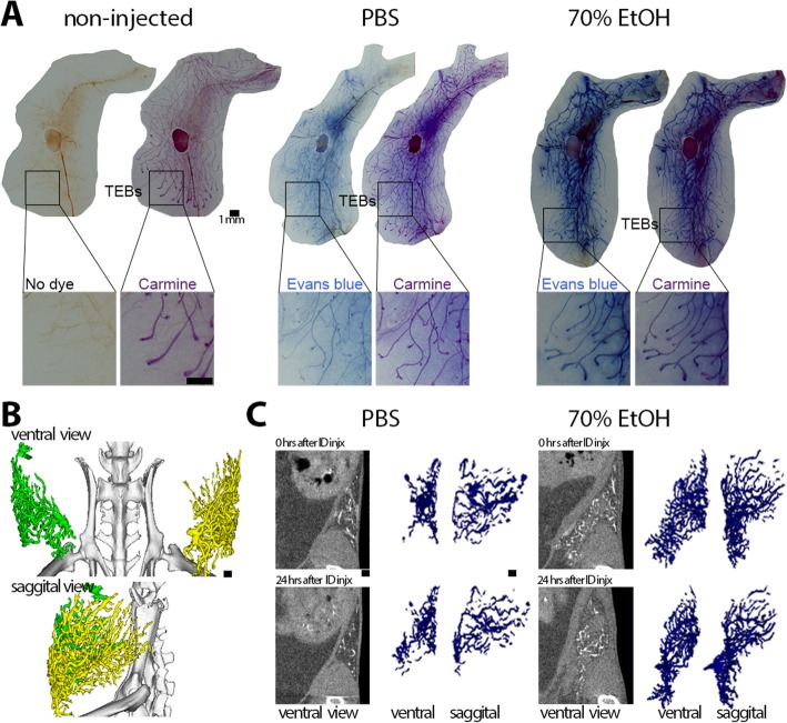
Fig. 1.
Feasibility of ductal tree filling and in vivo imaging of mammary glands with a 70% ethanol-containing solution. a Dual stain on whole-mount preparation of abdominal glands injected with Evans blue-containing solution of PBS or 70% EtOH. Evans blue serves to track injected solution within the lumen of the ductal tree, and carmine alum stains epithelial cells of the ductal tree. b Tantalum-based contrast agent-containing solution in PBS was sequentially injected within 15 min in the left abdominal (#4) and right abdominal gland (#9); 14-min high-resolution microCT scan was acquired 24 h after ID injection and processed for 3D image reconstruction. c Longitudinal 2-min standard microCT scans were acquired from independent animals whose abdominal glands were injected with tantalum-based contrast agent-containing solution of PBS or 70% EtOH. Different angle views and time points of the same representative glands are shown. Voxels with signal intensities from − 500 to 500 Hounsfield units in original CT slices were selected for volume rendition of diffused contrast agent. Scale bars indicate 1 mm in image panels at different magnification

