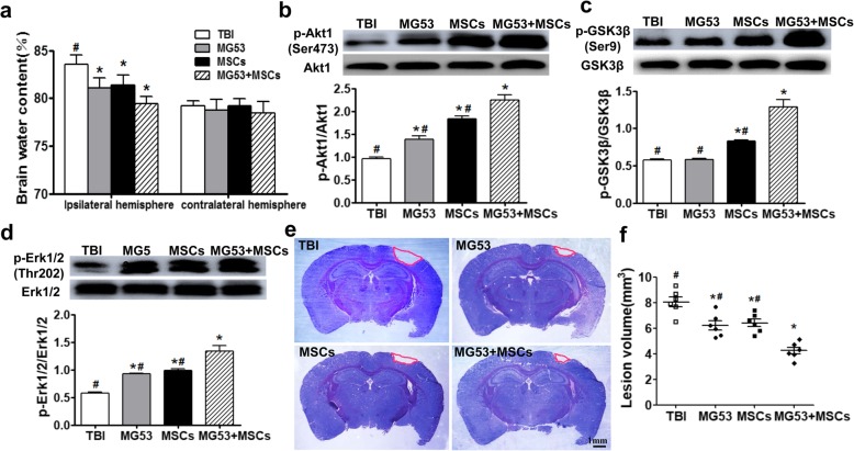
Fig. 4.
rhMG53 and hUC-MSCs decrease brain edema and reduce brain lesion volume after TBI by activating PI3K/Akt-GSK3β and ERK signaling. a Percentage of brain water content between ipsilateral hemisphere and contralateral hemisphere at day 3 post-TBI. Representative immunoblots and statistical analysis for phosphorylated and total Akt1 (b), GSK3β (c), and ERK 1/2 (d) in lysates from the injured brain tissues 3 days after TBI. e Cresyl violet staining of brain sections at 28 days post-TBI. Injured areas lack staining and are circled in red. Scale bar = 1 mm. f Quantification of lesion volume from the Cresyl violet-stained brains. Data for all graphs were presented as mean ± SEM. n = 6 per group. *p < 0.05, compared with TBI group; #p < 0.05, compared with MG53 + MSC group

