Fig. 1.
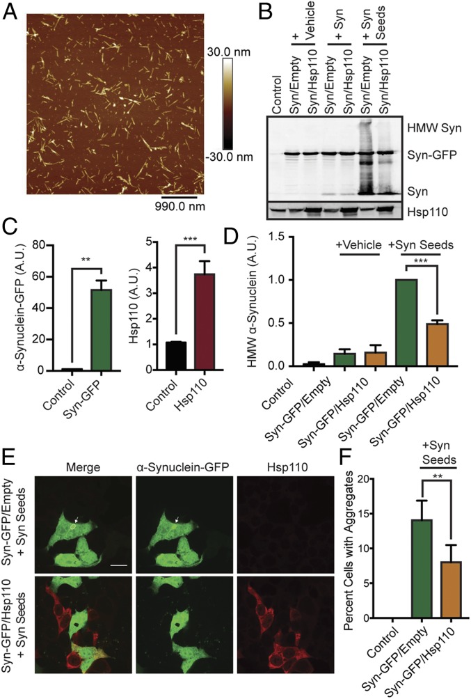
Hsp110 overexpression in mammalian cells alleviates α-synuclein aggregation. (A) Atomic force microscopy showing morphology of α-synuclein oligomers (Syn seeds). (B) Western blot of HEK293T cells overexpressing α-synuclein–GFP or both α-synuclein–GFP and Hsp110 treated with vehicle, α-synuclein fibrils (Syn), or α-synuclein oligomers (Syn seeds). HMW α-synuclein is indicative of internalization of seeds and intracellular α-synuclein aggregation. (C) Quantification of α-synuclein–GFP and Hsp110 overexpression in HEK293T Western blot, shown in B. (D) Quantification of HMW α-synuclein in HEK293T Western blot, shown in B. n = 3 experiments per condition. (E) Representative images of HEK293T cells transfected with α-synuclein–GFP (green) and Hsp110 (red) following α-synuclein aggregate addition. (Scale bar, 10 μm applies to all panels.) A GFP-positive α-synuclein aggregate is indicated by a white arrow. (F) Quantification of the percentage of GFP-positive HEK293T cells with intracellular GFP-positive aggregates templated from added α-synuclein seeds. Two-tailed Student t test: n = 6/condition; **P < 0.01; ***P < 0.001.

