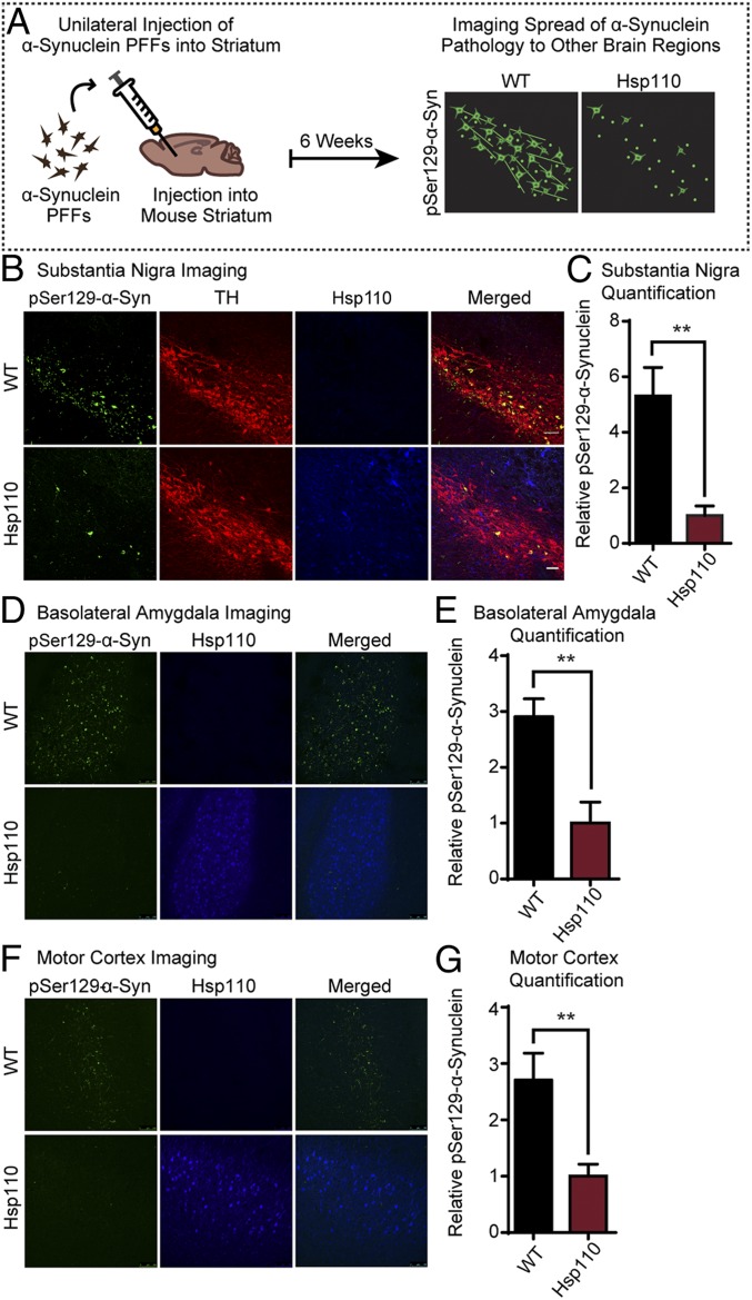
Fig. 5.
Transgenic expression of Hsp110 mitigates the spread of injected α-synuclein PFFs. (A) Schematic showing stereotaxic brain injections of α-synuclein PFFs and subsequent imaging. WT and Hsp110 mice were injected with α-synuclein PFFs into the striatum at 7 mo of age. After 6 wk, the mice were perfused, and the substantia nigra was imaged for the presence of pSer129–α-synuclein, indicating spread of pathology from the striatum. (B) Representative images of WT and Hsp110 mouse substantia nigra 6 wk postinjection, with pSer129–α-synuclein stained in green, tyrosine hydroxylase (TH) stained in red, and Hsp110 in blue. (C) Quantitation of pSer129–α-synuclein levels in WT and Hsp110 mouse substantia nigra. n = 6 mice/genotype; **P < 0.01, Student’s t test. (D) Representative images of WT and Hsp110 mouse basolateral amygdala 6 wk postinjection with pSer129–α-synuclein stained in green and Hsp110 in blue. (E) Quantitation of pSer129–α-synuclein levels in WT and Hsp110 mouse basolateral amygdala. n = 6 or 7 mice/genotype; **P < 0.01, Student’s t test. (F) Representative images of WT and Hsp110 mouse motor cortex 6 wk postinjection, with pSer129–α-synuclein stained in green and Hsp110 in blue. (G) Quantitation of pSer129–α-synuclein levels in WT and Hsp110 mouse motor cortex. (Scale bar, 50 μm, and applies to all panels in A, B, D, and F.) n = 6 or 7 mice/genotype; **P < 0.01, Student’s t test.

