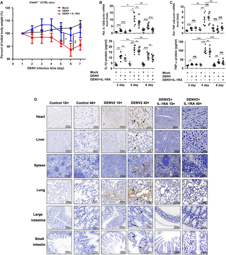
FIGURE 4.
Dengue virus (DENV) promotes interleukin (IL)-1β release in IFNAR–/– C57BL/6 mice. (A–D) IFNAR–/– C57BL/6 mice were intravenously injected with 300 μl of DENV2(NGC) suspension at a dose of 1 × 10⁶ PFU/mouse (n = 6), pretreated with intraperitoneal injection of 300 μl of PBS containing 2 μg of mouse IL-1RA at 90 min before the infection with DENV2(NGC) (1 × 10⁶ PFU/mouse) with the IL-1RA treatment repeated 4 days after the infection (n = 6) or injected with 300 μl of PBS only (control group, n = 6). Mice were euthanized 7 days after infection or PBS injection, and the tissues were collected. (A) Mice were weighed daily; body weight is expressed as the percentage of the initial weight. (B) Blood samples were collected at 2, 4, and 6 days postinfection. IL-1β mRNA in blood cells was determined by qRT-PCR (top), and IL-1β protein in the serum was measured by ELISA (bottom). Individual points represent the IL-1β value in each sample. (C) Blood samples were collected at 2, 4, and 6 days postinfection. TNF-α mRNA in blood cells was determined by qRT-PCR (top), and TNF-α protein in the serum was measured by ELISA (bottom). Individual points represent the TNF-α value in each sample. (D) Detection of IL-1β by immunohistochemistry in the heart, liver, spleen, lung, large intestine, and small intestine after DENV infection. Black arrows indicated the immunostaining of IL-1β. Mock: injection of the same volume of PBS. Data represent two independent experiments. Values are mean ± SEM; ns, not significant; ∗, ∗∗, ∗∗∗ indicate P-values less than 0.05, 0.01, and 0.001, respectively.

