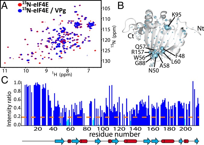
Fig. 3.
Interaction surface used by eIF4E to bind VPg. (A) 1H-15N HSQC of 15N-labeled eIF4E (50 μM) in the absence (red) or presence (blue) of 3-molar excess VPg. (B) Broadening and CSPs mapped onto the apo-eIF4E structure (PDB ID code 2GPQ) depicted as light blue balls. (C) Per residue plot of backbone amide line broadening of 50 μM eIF4E in response to binding of VPgΔ37 (extracted from A). Residues that undergo line broadening below the dashed line are in cyan.

