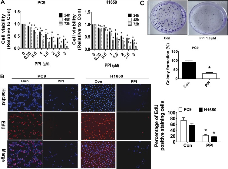
Figure 1.
PPI inhibited growth of NSCLC cells. (A) PC9 and H1650 cells were stimulated with different concentrations of PPI for up to 72 h. The cells were collected and processed for MTT assay as described in the Materials and Methods section. (B) PC9 and H1650 cells were treated with PPI (1.6 μM) for 24 h, followed by processing for measuring the cell growth by the EdU DNA cell proliferation kit as described in the Materials and Methods section. (C) PC9 cells (5×10² cells/6-well plate) were seeded in 6-well plates and treated with PPI (1.6 μM) at 37 °C in 5% humidified CO2 for up to 9 days. After that, the cells were fixed with 4% paraformaldehyde (Sigma-Aldrich) and stained by 0.1% crystal violet, and the visible colonies were counted under a microscope as described in the Materials and Methods section. Values are given as the mean±SD, from three independent experiments performed in triplicate. *Indicates significant difference as compared to the untreated control group (P<0.05).

