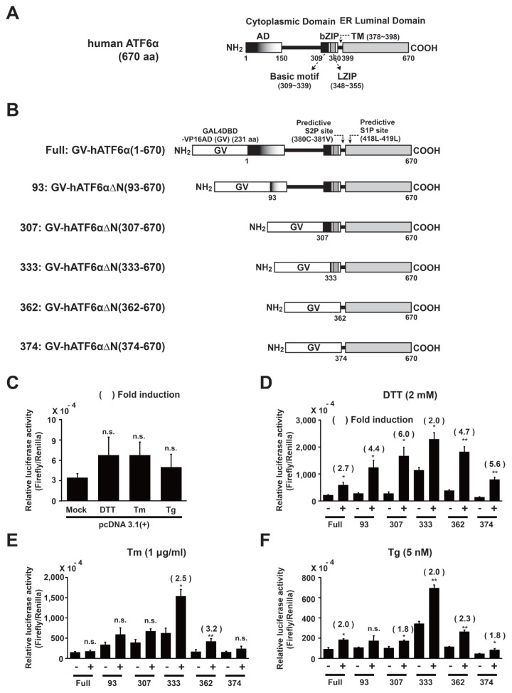
Fig. 1. Selection of a GV-ATF6α fusion variant to measure regulated intermembrane proteolysis of ATF6α in HLR cell line during ER stress.
(A) Predicted domain structure of human ATF6α full length. (B) Schematic diagrams of fusion proteins expressed from pCMV-GAL4DBD-VP16AD(GV)-ATF6α full or deletion variants as described in Materials and Methods section. (C–F) HLR cells were transfected with each expressing plasmid, including pcDNA 3.1(+) and pRL-CMV as internal controls. DTT (2 mM), Tm (1 μg/ml), or Tg (5 nM) was used for treatment for 12 h. Firefly luciferase and Renilla luciferase activities were measured as described in Materials and Methods section. Results are given as absolute values of firefly luciferase normalized against Renilla luciferase activities in each cell lysate. The number of parenthesis indicates average fold induction relative to the untreated group. Data are expressed as mean ± SD of three independent experiments. *P < 0.05, **P < 0.01 (untreated group vs each treated group); n.s., not significant.

