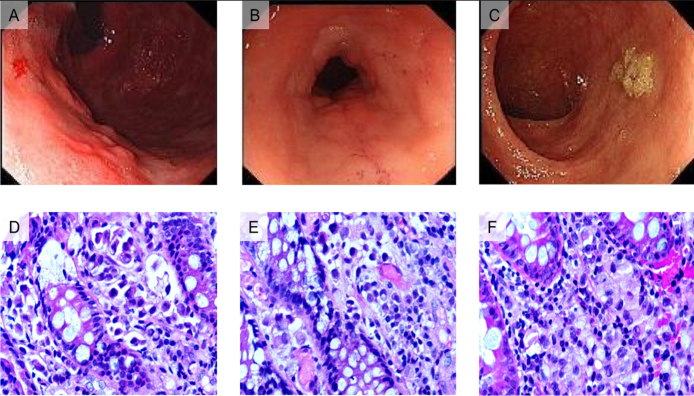
Figure 1.
Endoscopic views and pathological staining of gastric and colon specimens from two patients.
A) The gastroscopic view of the first patient found a local protuberant lesion on the body of the stomach and obvious thickening of the mucosa of the gastroesophageal angle. B) A colon stenosis was identified by colonoscopy in the first patient. C) The colonoscopic features of the second patient showed a flat bulging. D) Numerous signet ring cells were observed in the hematoxylin and eosin (HE) staining of the gastric specimen obtained from the first patient. E) HE staining of the colon specimen showing strands of invasive cells in the mucosa. F) HE staining of the colon specimen obtained from the second patient showing strands of invasive cells in the mucosa.

