
Figure 3a:
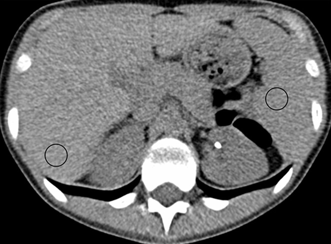
(a, b) Abdominal CT images (window/level = 50/330 HU) in a 16-year-old girl acquired with the institution’s dose class 3 protocol (CT dose index: 1.56 mGy, 120 kV). Images are reconstructed in the transaxial plane with (a) Siemens weighted filtered back projection (kernel B31f) and (b) convolutional neural network processing. Hounsfield unit mean and noise power measured by HU standard deviation in the region of interest (circle) for liver and spleen were as follows: liver, HUmean = 62.5, HUSD = 17.6; spleen: HUmean = 51.3, HUSD = 19.0 in a; and liver: HUmean = 62.2, HUSD = 11.5; spleen: HUmean = 51.1, HUSD = 12.6 in b.