
Figure 4a:
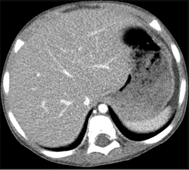
(a–c) Abdominal CT images in a 3-year-old girl acquired with the institution’s dose class 2 protocol (CT dose index: 0.50 mGy, 70 kV). Images are reconstructed in the transaxial plane with (a) Siemens weighted filtered back projection (kernel Br44d), (b) advanced modeled iterative reconstruction (kernel Br44d\3), and (c) convolutional neural network processing.