Figure 1.
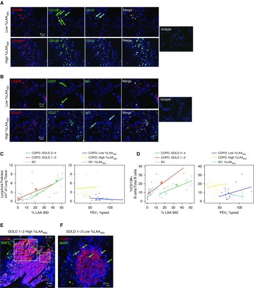
Increases in B cell–adaptive immune responses are associated with the extent of emphysema and not with airflow limitation. (A) Triple immunofluorescence staining for CD45R (B-cell marker), CD138 (plasma-cell marker), CD10 (immature B-cell and follicle center B-cell [centrocyte] marker), and the merge panel. The green arrows indicate CD138+ B cells, and cyan arrows indicate CD10+ B cells. (B) Triple immunofluorescence staining for CD45R, CD27 (memory B cells), IgGs, and the merge panel. The green arrows indicate CD27+ B cells, and the cyan arrows indicate IgG+ B cells. Isotype control merge figures are shown on the right of both A and B. (C and D) Stratified graphs are presented for the association of low-attenuation areas below a threshold of −950 Hounsfield units (%LAA950) and FEV1% predicted (FEV1%pred) with (C) the number of lymphoid follicles (LFs)/cm2 of lung tissue and (D) the %CD138+ B cells/total B cells. For each graph, the relationship of the parameter of interest with %LAA950 within different Global Initiative for Obstructive Lung Disease (GOLD) stages (1–2 vs. 3–4) is shown in the left panel, and the relationship with FEV1%pred within different levels of emphysema is shown in the right panel. SC = smokers without chronic obstructive pulmonary disease (COPD). (E and F) Double-immunofluorescence pictures of formalin-fixed paraffin-embedded lung sections from 1) a patient with GOLD 1–2 COPD and severe emphysema (E), showing robust BAFF (B-cell activation factor of the TNF family) staining in most of the alveolar cells, LF B cells, and parenchymal B cells; and 2) a patient with GOLD 1–2 COPD and low emphysema (F), showing fewer BAFF+ alveolar cells, B cells within the LF, and parenchymal B cells. In E, the inset shows a detail of an LF, with the great majority of B cells expressing BAFF. The green arrows indicate BAFF+ B cells and alveolar cells.

