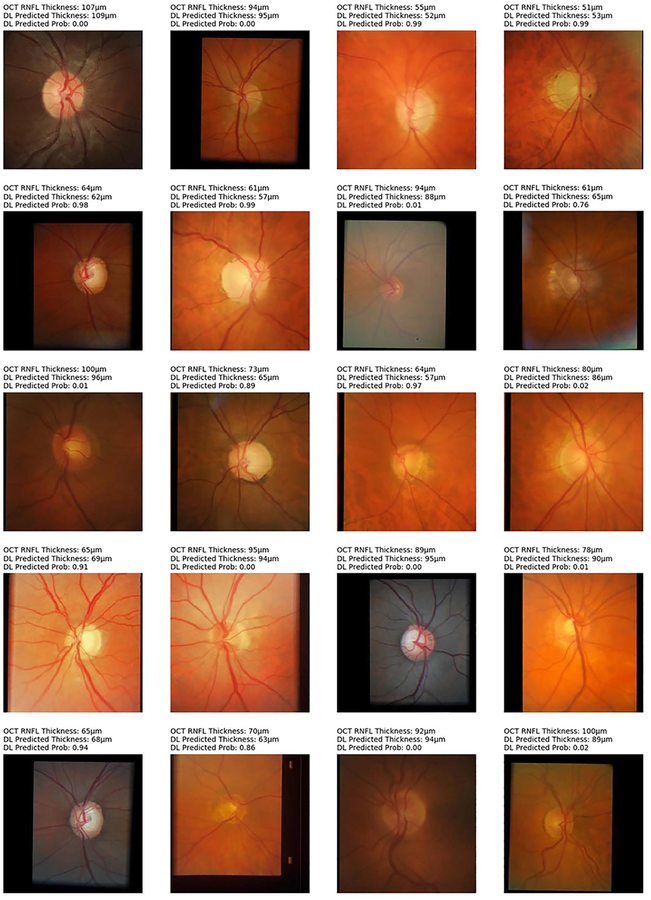
Figure 6.
Random examples of optic disc photographs that were correctly classified according to the reference classification of the Spectralis spectral domain-optical coherence tomography (OCT) normative database for average retinal nerve fiber layer thickness (RNFL). Above each photo is shown the OCT average thickness measurement, the deep learning (DL) prediction of average RNFL thickness from the optic disc photograph, and the probability of abnormality estimated by the DL algorithm.

