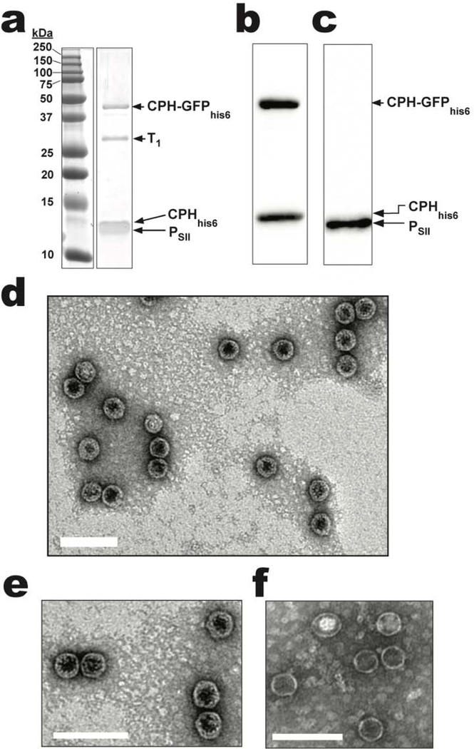
Figure 2.
CPH-GFPhis6 incorporation into CPHhis6-T1-PSII shells. a) SDS-PAGE analysis of purified CPHhis6-T1-PSII/CPH-GFPhis6 shells (see Figure S1 for the uncropped SDS-PAGE image). b) Anti-his or c) anti-strep western blots of purified shells from (a). d) TEM micrograph of purified CPHhis6-T1-PsII/CPH-GFPhis6 shells. e) Further magnified TEM micrograph of CPHhis6-T1-PSII/CPH-GFPhis6 shells. f) For comparison, magnified TEM micrograph of CPH-T1-PsII shells without GFP cargo from Figure 1e. Scale bars are 100 nm.

