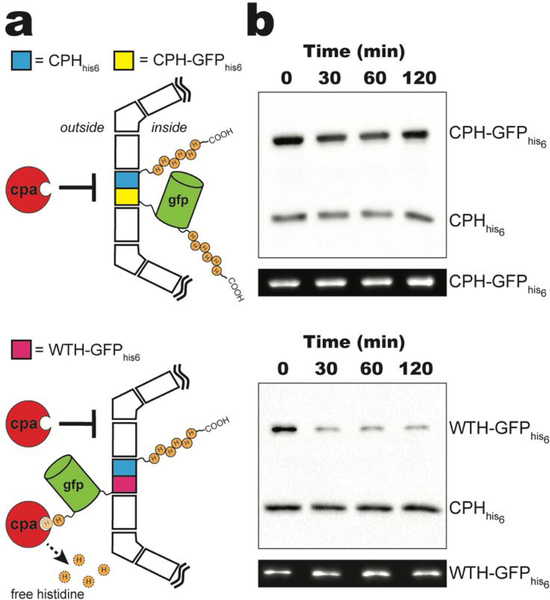
Figure 3.
In vitro protease protection assay. a) Schematic of carboxypeptidase A (CPA) protection assay for purified CPHhis6-T1-PSII shells incorporating CPH-GFPhis6 (top) or WTH-GFPhis6 (bottom). For clarity, only one CPHhis6 (cyan) and CPH-GFPhis6 (yellow) or WTH-GFPhis6 (pink) is shown per shell. b) Western blot of a CPA time course of purified CPHhis6-T1-PSII shells incorporating CPH-GFPhis6 (top) or WTH-GFPhis6 (bottom) using anti-his antibodies. Representative in-blot intrinsic GFP fluorescence is shown below each western blot. See Figure S3 for uncropped fluorescence images.

