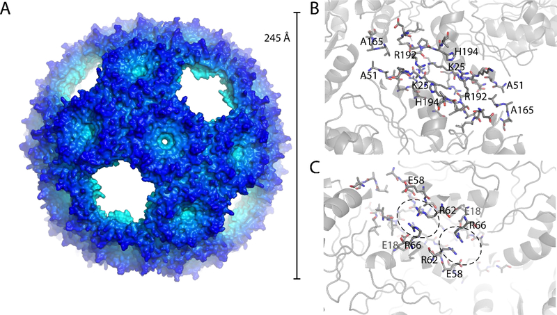
Figure 3. Structure of the BMC-H2 shell.
A: Surface representation of the crystal structure of the BMC-H2 T=4 icosahedral shell. B: View on the trimer-trimer interface from the outside with interacting residues shown as sticks. C: View from the inside, interacting arginine residues are highlighted as well as charge complementing glutamic acid residues.

