
Figure 4. IDO treated DCs suppress antigen specific T cell proliferation, and suppression is active enzyme dependent.
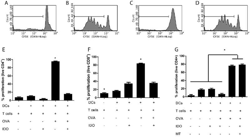
Dendritic cells were incubated with IDO in the presence or absence of 1-methyl tryptophan (MT) for 24 h, then washed and pulsed with the corresponding ovalbumin peptide (OVA) for 3 h. Dendritic cells were then washed and co-cultured with CD4+ or CD8+ CFSE labeled T cells isolated from OT-II and OT-I mice respectively, for 4 d. T cell proliferation was quantified through CFSE dilution via flow cytometry. Representative histograms for live CD4+ T cells, stimulated with: A. Untreated DCs, B. DCs pulsed with OVA, C. IDO-treated DCs pulsed with OVA, D. IDO-MT-treated DCs pulsed with OVA. T cell proliferation was quantified for E. CD4+ and F. CD8+ populations where “+” and “−” represent presence or absence of a specific component. G. CD4+ T cell proliferation assay with the introduction of MT during IDO treatment of DCs. Shown is the mean ± SEM of three separate experiments, each conducted in triplicate. * denotes pair-wise significant differences (p ≤ 0.05) from all other groups by ANOVA, with Tukey’s post-hoc test.