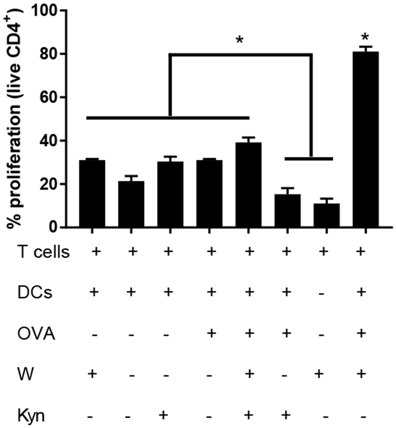
Figure 5. Tryptophan depletion and kynurenine accumulation combine to maximally suppress antigen specific proliferation.

Dendritic cells were cultured in tryptophan (W) -free media supplemented with kynurenine (Kyn) or relevant controls for 24 h then pulsed with ovalbumin peptide (OVA) for an additional 3 h. Cells were washed and co-cultured with CD4+ CFSE labeled T cells isolated from OT-II mice for 4 d. “+” and “−” denote the presence or absence of a particular component during assay. Proliferation of live T cells was quantified through CFSE dilution via flow cytometry. Shown is the mean ± SEM of three separate experiments, each conducted in triplicate. * denotes pair-wise significant differences (p ≤ 0.05) from all other groups, while * above brackets indicates pair-wise significant differences (p ≤ 0.05) from indicated groups by ANOVA, with Tukey’s post-hoc test.
