Figure 4.
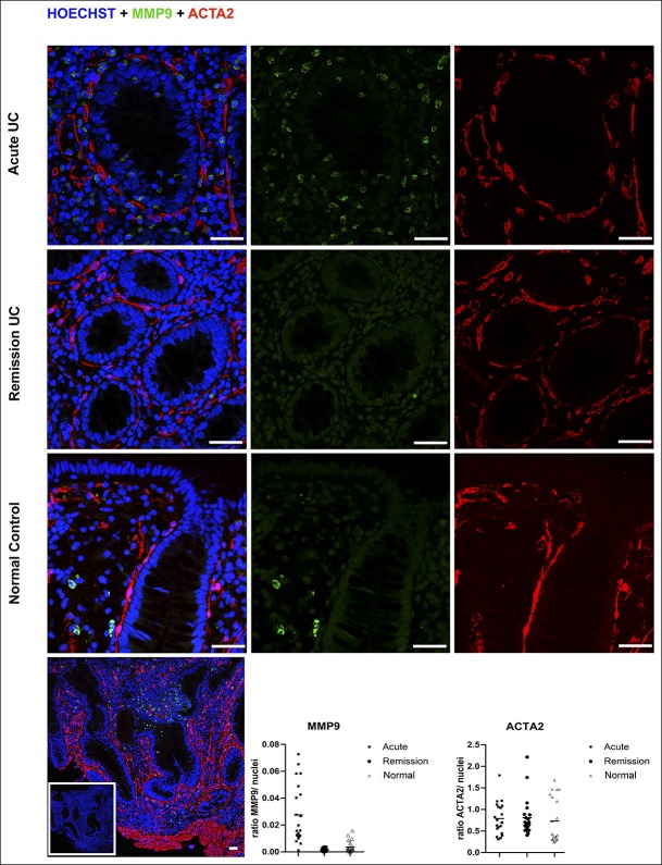
Immunostaining of MMP9 and ACTA2. Antibodies directed at MMP9 and ACTA2 had the highest upregulated and downregulated gene patterns in our study with enhanced antibody scores. Panels show representative immunostaining of formalin-fixed, paraffin-embedded colonic endoscopic biopsies from acute UC, remission UC, and a healthy control group at ×400 magnification. Scale bars = 35 μm. Dual staining with monoclonal rabbit antibody against MMP9 ([1/400], Cell Signaling Technologies) and monoclonal mouse antibody against ACTA2/SMA (DAKO, [0.35 μg/mL]) were used. Cell nuclei are stained with Hoechst (blue). Positive cytosolic staining of stromal cells was observed for MMP9 (green), whereas a membranous/cytoplasmic signal was seen in ACTA2-positive cells (red). A significant decrease (P < 0.05) of MMP9 was seen comparing acute UC with remission UC or normal controls, but not for ACTA2. Further antibody details are given in Supplemental Table 1(see Supplementary Digital Content, http://links.lww.com/CTG/A103). A negative control is shown in the white frame for acute UC (bottom left panel). ACTA2, actin alpha 2; UC, ulcerative colitis.

