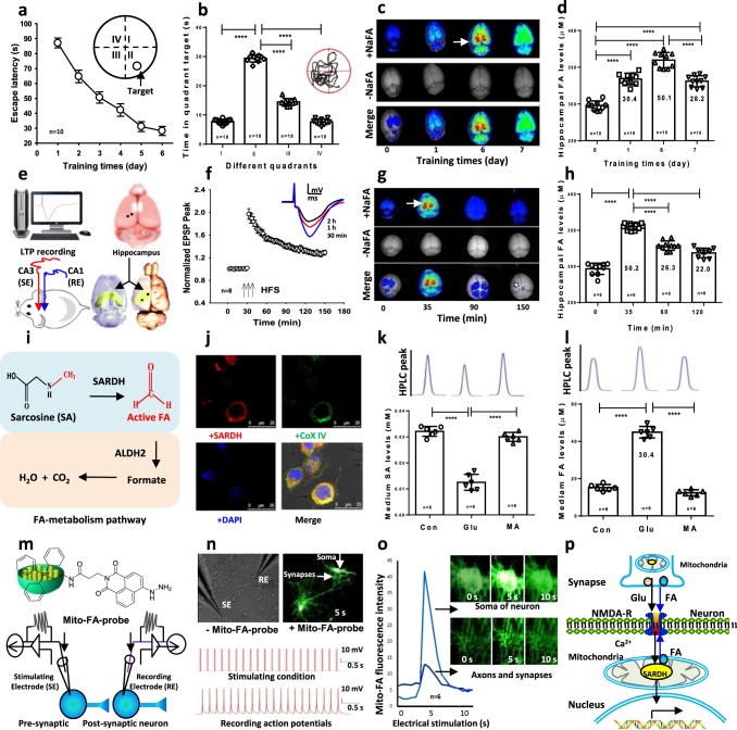
Fig. 1. Spatial learning elicits formaldehyde generation.
a, b Spatial learning and memory in wild-type SD rats trained in the MWM (n = 10 per group). c Brain formaldehyde fluorescence revealed by the in vivo imaging system. NaFA: a fluorescence probe of free formaldehyde, n = 3. d Hippocampal formaldehyde (FA) levels detected by Fluo-HPLC (n = 10). e An in vivo LTP recording in CA1 from Schaffer collateral stimulation and 3D views of the hippocampus (yellow). SE stimulating electrode, RE recording electrode. f Late-LTP (L-LTP) formation in vivo. n = 8, HFS high-frequency stimulation. g Brain formaldehyde revealed by the in vivo imaging system, n = 3. h Hippocampal formaldehyde levels detected by Fluo-HPLC (n = 6). i The pathway of formaldehyde metabolism. SARDH sarcosine dehydrogenase, ALDH2 aldehyde dehydrogenase. j Colocalization of SARDH (red) and the mitochondrial marker- Cox IV (green) in the cultured hippocampal neurons. DAPI: blue, a nuclear dye. k, l The cultured medium sarcosine and formaldehyde levels detected by Fluo-HPLC. SA sarcosine, MA methoxyacetic acid, an inhibitor of SARDH, n = 6. m Intracellular infusion of the mitochondrial formaldehyde probe (mito-FA-probe). n A train of 20 pulses in the presynaptic neurons induced multiple action potentials in the postsynaptic neurons, n = 8. o The active formaldehyde generated from mitochondria in the axon, synapse and soma of the cultured neurons imaged by the mito-FA probe, n = 6. p The model of endogenous formaldehyde-enhanced memory formation. The data are expressed as the mean ± standard error (s.e.m.). ***p < 0.001; ****p < 0.0001.

