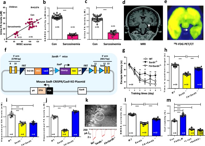
Fig. 5. SARDH mutation-induced formaldehyde deficiency causes amnesia.
a Positive relationship between urine formaldehyde (FA) levels and Wechsler Intelligence Scale for Children (WISC) scores in 11 pediatric sarcosinemia patients and 31 healthy children. b Blood SARDH activity analyzed by a human SARDH kit (p < 0.01). c Urine formaldehyde concentrations detected by Fluo-HPLC (p < 0.01). d Right hippocampal atrophy revealed by MRI. e Impaired glucose metabolism in the right hippocampus revealed by 18F-FDG PET/CT. f The scheme for generation of Sardh−/− mice. g After 6 days of MWM training, repeated measures two-way ANOVA revealed a difference in group: (F(2, 27) = 16.38, p < 0.001), training time (F(5, 152) = 72.54, p < 0.001), and a group × time interaction (F(10, 152) = 2.45, p < 0.001). Post hoc tests showed that the mean escape latency values for SARDH−/− mice were a significant longer than control (wild-type mice) on day 3 (F(2, 27) = 2.41, p = 0.001), day 4 (F(2, 27) = 4.57, p = 0.005), day 5 (F(2, 27) = 6.39, p = 0.003), and day 6 (F(2, 27) = 6.43, p = 0.002); while there was no statistically significant difference in escape latency between SARDH−/− mice with 200 μM formaldehyde injection and control (p > 0.05; n = 10 mice per group). h SARDH−/− mice with formaldehyde infusion reversed the reduced time in target quadrant of these mice without formaldehyde treatment (p < 0.001). i Hippocampal sarcosine levels detected by a mouse sarcosine kit, n = 6. j Hippocampal formaldehyde levels detected by Fluo-HPLC, n = 6. k, l NMDA currents recording in the brain slices of Sardh−/− mice and formaldehyde-injected Sardh−/− mice, n = 10−12. m The effects of formaldehyde scavengers- l-cys (300 μM) and NaHSO3 (300 μM) on the cell viability of the cultured hippocampal neurons detected by CCK-8 kit, n = 6. Data are expressed as the mean ± standard error (s.e.m.). ***p < 0.001; ****p < 0.0001.

